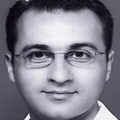
Filip A. Risteski
Work place: Skopje City General Hospital, Pariska B.B., 1000 Skopje, Macedonia
E-mail: risteskifilip@bolnica.org.mk
Website:
Research Interests: Physics, Computational Physics
Biography
Filip A. Risteski was awarded the degree of Physician at the Faculty of Medicine, St. Cyril and Methodious University in Skopje, Macedonia in 2005. Following the completion of the degree of Physician, Dr. Risteski earned the position of Medical Specialist in Radiology from the Radiology Institute at the Faculty of Medicine, St. Cyril and Methodious University in Skopje, Macedonia. Dr. Risteski is now with the Skopje City General Hospital, Skopje, Macedonia since 2011. During the years 2002-2009 Dr. Risteski has been an active contributor of scientific articles in the field of applied radiology, and he is currently a member of the Macedonian Medical Association, the Macedonian Radiologists Association, the European Society of Radiology, the European Society of Skeletal Radiology and the Radiological Society of North America.
Author Articles
A Novel Approach to T2-Weighted MRI Filtering: The Classic-Curvature and the Signal Resilient to Interpolation Filter Masks
By Carlo Ciulla Farouk Yahaya Edmund Adomako Ustijana Rechkoska Shikoska Grace Agyapong Dimitar Veljanovski Filip A. Risteski
DOI: https://doi.org/10.5815/ijieeb.2016.01.01, Pub. Date: 8 Jan. 2016
This paper presents a novel and unreported approach developed to filter T2-weighetd Magnetic Resonance Imaging (MRI). The MRI data is fitted with a parametric bivariate cubic Lagrange polynomial, which is used as the model function to build the continuum into the discrete samples of the two-dimensional MRI images. On the basis of the aforementioned model function, the Classic-Curvature (CC) and the Signal Resilient to Interpolation (SRI) images are calculated and they are used as filter masks to convolve the two-dimensional MRI images of the pathological human brain. The pathologies are human brain tumors. The result of the convolution provides with filtered T2-weighted MRI images. It is found that filtering with the CC and the SRI provides with reliable and faithful reproduction of the human brain tumors. The validity of filtering the T2-weighted MRI for the quest of supplemental information about the tumors is also found positive.
[...] Read more.Applied Computational Engineering in Magnetic Resonance Imaging: A Tumor Case Study
By Carlo Ciulla Dijana Capeska Bogatinoska Filip A. Risteski Dimitar Veljanovski
DOI: https://doi.org/10.5815/ijigsp.2014.07.01, Pub. Date: 8 Jun. 2014
This paper solves the biomedical engineering problem of the extraction of complementary and/or additional information related to the depths of the anatomical structures of the human brain tumor imaged with Magnetic Resonance Imaging (MRI). The combined calculation of the signal resilient to interpolation and the Intensity-Curvature Functional provides with the complementary and/or additional information. The steps to undertake for the calculation of the signal resilient to interpolation are: (i) fitting a polynomial function to the signal, (ii) the calculation of the classic-curvature of the signal, (iii) the calculation of the Intensity-Curvature term before interpolation of the signal, (iv) the calculation of the Intensity-Curvature term after interpolation of the signal, (v) the solution of the equation of the two aforementioned Intensity-Curvature terms of the signal provides with the signal resilient to interpolation. The Intensity-Curvature Functional is the result of the ratio between the two Intensity-Curvature terms before and after interpolation. Because of the fact that the signal resilient to interpolation and the Intensity-Curvature Functional are derived through the process of re-sampling the original signal, it is possible to obtain an immense number of images from the original MRI signal. This paper shows the combined use of the signal resilient to interpolation and the Intensity-Curvature Functional in diagnostic settings when evaluating a tumor imaged with MRI. Additionally, the Intensity-Curvature Functional can identify the tumor contour line.
[...] Read more.Computational Intelligence in Magnetic Resonance Imaging of the Human Brain: The Classic-Curvature and the Intensity-Curvature Functional in a Tumor Case Study
By Carlo Ciulla Dijana Capeska Bogatinoska Filip A. Risteski Dimitar Veljanovski
DOI: https://doi.org/10.5815/ijieeb.2014.02.01, Pub. Date: 8 Apr. 2014
This research solves the computational intelligence problem of devising two mathematical engineering tools called Classic-Curvature and Intensity-Curvature Functional. It is possible to calculate the two mathematical engineering tools from any model polynomial function which embeds the property of second-order differentiability. This work presents results obtained with bivariate and trivariate cubic Lagrange polynomials. The use of the Classic-Curvature and the Intensity-Curvature Functional can add complementary information in medical imaging, specifically in Magnetic Resonance Imaging (MRI) of the human brain.
[...] Read more.Other Articles
Subscribe to receive issue release notifications and newsletters from MECS Press journals